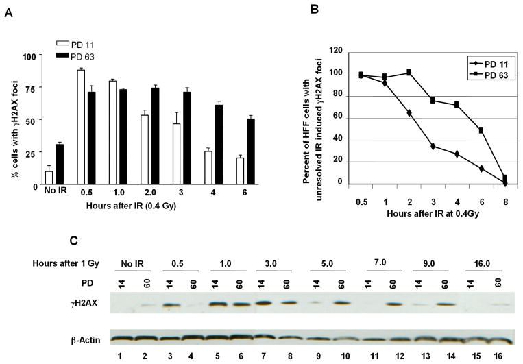
Fig. 2. Kinetics of γH2AX focus formation and resolution are delayed in cells with short telomeres.
A, Early (PD 11) and late-passage (PD 63) HFF were irradiated with 0.4 Gy and processed for immunofluorescence using anti-γH2AX antibodies at the indicated time post-IR. A total of 100 cells were analyzed, cells with 3 or more foci were considered γH2AX-positive. B, time course of γH2AX resolution, the percentage of cells with unresolved γH2AX IR-induced foci was assessed in early and late-passage at the indicated time following irradiation with 0.4 Gy. C, Early and late-passage HHF were irradiated with 1 Gy and harvested at the indicated time points post-IR for histone protein extraction. Phosphorylated H2AX protein levels were assessed using anti-γH2AX antibodies.

