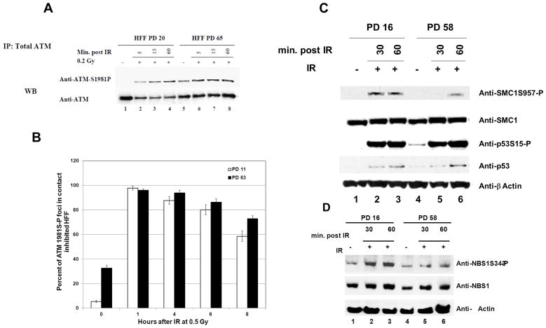
Fig. 4. Delayed phosphorylation kinetics of chromatin bound proteins.
A, ATM phosphorylation kinetics post-IR is similar in early and late-passage HFF. HFF at PD 20 and 65 were irradiated with 0.2 Gy. Cells were harvested at the indicated time points post-IR and whole–cell extracts were prepared. Cleared supernatants were immunoprecipitated with anti-ATM, and the samples were resolved by gel electrophoresis and immunoblotted with anti-total ATM and anti-ATM 1981S-P. B, ATM1981S-P foci formation kinetics are similar in early and late-passage HFF. HFF at PD 11 and 63 were irradiated with 0.5 Gy and the kinetics of ATMS1981-P foci formation were assessed at the indicated time points post-IR. A total of 100 cells per time point were analyzed, cells with 3 or more foci were considered ATMS1981-P positive. The values shown are the mean +/− S.D. of 5 fields analyzed of 100 cells each. C and D, HFF at PD 16 and 58 were irradiated with 10 Gy and harvested at the indicated time points for immunoblotting. Total and phosphorylated p53 SMC1 and NBS1 were assessed.

