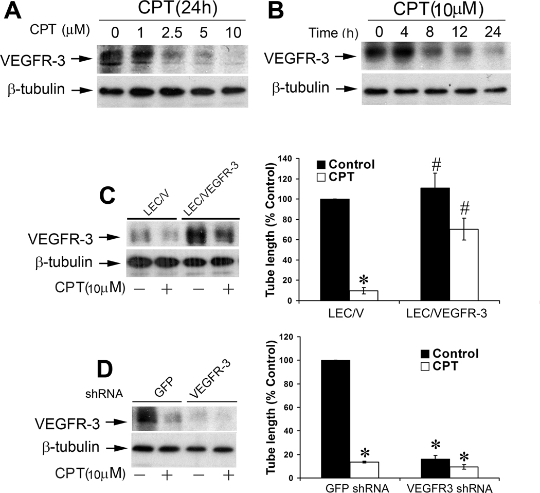
Figure 2.
CPT inhibition of LEC tube formation is associated with suppressing VEGFR-3 protein expression. A, B, CPT inhibited protein expression of VEGFR-3 in a concentration- and time-dependent manner. LECs, treated with CPT (0–10 µM) for 24 h (A) or CPT (10 µM) for 0–24 h (B), were harvested and subjected to Western blot analysis with antibodies to VEGFR-3. β-tubulin was used as a loading control. C, Overexpression of VEGFR-3 partially prevented CPT inhibition of LEC tube formation. LEC/V (control) and LEC/VEGFR-3 cells were treated with CPT (10 µM) for 24 h, followed by Western blot analysis with indicated antibodies (Left panel), or tube formation assay (Right panel), as described in “Materials and Methods”. Quantitative results of tube formation are shown as mean ± SD (n = 3). *P < 0.05, difference vs. control group; #P < 0.05, difference vs. LEC/V group. D, Lentiviral shRNA to VEGFR-3, but not GFP, downregulated VEGFR-3 protein expression in LECs, as detected by Western blotting (Left panel). LECs, infected with lentiviral shRNAs to VEGFR-3 and GFP (control), respectively, were treated with CPT (10 µM) for 24 h, followed by tube formation assay (Right panel), as described in “Materials and Methods”. Quantitative results of tube formation are shown as mean ± SD (n = 3). * P < 0.05, difference vs. GFP shRNA control group.

