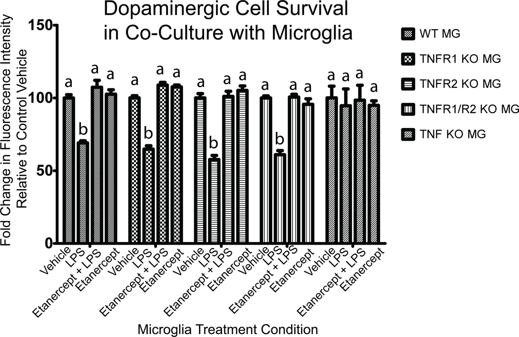
Figure 10. LPS-induced TNF secretion by primary microglia but not autocrine TNF signaling is required for microglial toxicity on dopaminergic cells.
Primary microglia from postnatal TNF-null, TNFR1-null, TNFR2-null, or TNFR1/R2 null mice were put in co-culture with neurally differentiated dopaminergic MN9D cells. Twenty-four hours after stimulation of co-cultures with LPS (1 ug/mL or saline vehicle in the presence or absence of the TNF decoy receptor etanercept (200 ng/mL) MN9D cell survival was assessed by quantifying the number of tyrosine hydroxylase (TH)-positive cells as described under Methods. A two-way ANOVA and Bonferroni's post hoc test. Columns with different letters are statistically significantly different from each other at p<0.05.

