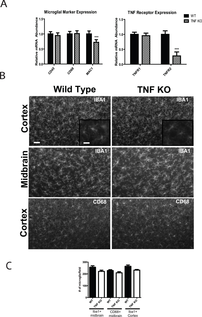
Figure 3. TNF signaling is not required for expansion of brain microglia but is critical for expression of Mac-1 and TNFR2 in adult brain.
(A) Brains (n = 4 mice/genotype) were rapidly removed and microdissected. Total RNA was extracted and reverse transcribed into cDNA for real-time PCR analysis of microglial surface markers or TNF receptors. Expression of CD45, CD68, and TNFR1 mRNA was not significantly different between genotypes but a significant reduction in expression of MAC1 and TNFR2 mRNAs was detectable in brain of adult TNF-null mice. Unpaired t-test, *p < 0.05, ***p < 0.001. (B) Immunohistochemical analysis of microglial markers Iba-1 and CD68 in midbrain or cortex of adult WT and TNF-null mice. Scale bar = 100 µm in low magnification images and 50 µm in the inset images. (C) Quantification of Iba-1 and CD68-positive cells per field in midbrain and cortex (See Methods) revealed no significant differences between genotypes.

