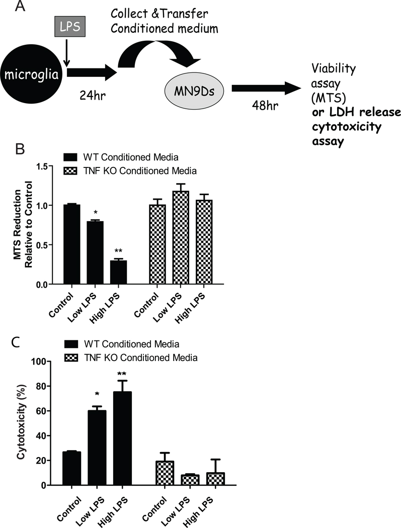
Figure 9. TNF-null microglia display reduced cytotoxicity on differentiated dopaminergic MN9D cells.
Schematic of the target-effector assay (A); Primary microglia isolated from wild type or TNF-null postnatal day 3–5 pups were plated and treated for 24 hrs with low (10 ng/mL) or high (1 µg/mL) LPS. Conditioned media (CM) from resting or treated microglia were collected and transferred directly onto cultures of differentiated dopaminergic MN9D cells for 48 hrs. Metabolic activity of MN9D dopaminergic cell line was assayed using an MTS reduction assay. Two-way ANOVA with Bonferroni’s post hoc test. *p < 0.05, **p < 0.01 (B); Cytotoxicity of CM on MN9D cells was assayed using an LDH release assay. Two-way ANOVA with Bonferroni’s post hoc test. *p < 0.05, **p < 0.01 (C).

