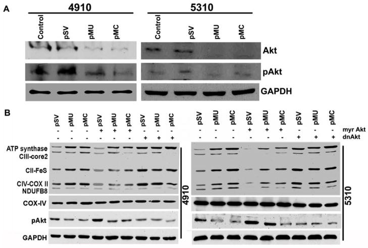
Figure 3. pMU and pMC mediate Akt-dependent OXPHOS activation in glioma.
(A) Cell lysates were assessed for pAkt and Akt levels by Western blotting. (B) Glioma cells were transfected with pSV, pMU or pMC for 48 hrs followed by transfection with myr-Akt or dnAkt and cultured for another 24 hrs. Cells were harvested and lysed, and fractions of mitochondrial and total cell lysates were collected separately. These fractions were used for Western blot analysis of OXPHOS complexes and pAkt. COX-IV indicated purity of mitochondrial fractions. GAPDH served as a loading control for total cell lysates. Results are representative of three independent experiments.

