
Figure 2.
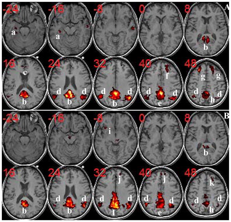
Group DMN components of normal controls (A) and aMCI (B) with corrected p<0.05. Resting-state activities for normal controls are found in: a=Left HC/PHG/FG; b=PCC/RC/Precuneus; c=ACC; d=AG; e=Precuneus/MCC; f=Right LPFC; g=LPFC; h=Precuneus. For aMCI subjects, there are no activities in left HC/PHG/FG (a) and left LPFC (see k=right LPFC). On the other hand, some activities extended into MCC (l=Precuneus/PCC/MCC), in MPFC (j), and in right insular cortext (i) are observed for aMCI. (The t-score range is from 3.5 to 16. ACC=Anterior Cingulate Cortex; AG=Angular Gyrus; FG=Fusiform Gyrus; HC=Hippocampus; LPFC=Lateral Prefrontal Cortex; MCC=Middle Cingulate Cortex; MPFC=Medial Prefrontal Cortex; PCC=Posterior Cingulate Cortex; PHG=Parahippocampal Gyrus; RC=Retrosplenial Cortex.)