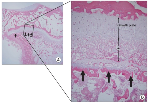
Fig. 3.
A histologic section of a compressed specimen harvested at four weeks after surgery showed newly formed trabecular bones that were interconnected haphazardly (asterisk) and transversely connected trabecular bone (arrows), the latter of which corresponded with the highly attenuated line observed in the contrast-enhanced micro-computed tomography (A: × 10, B: H&E, × 100).

