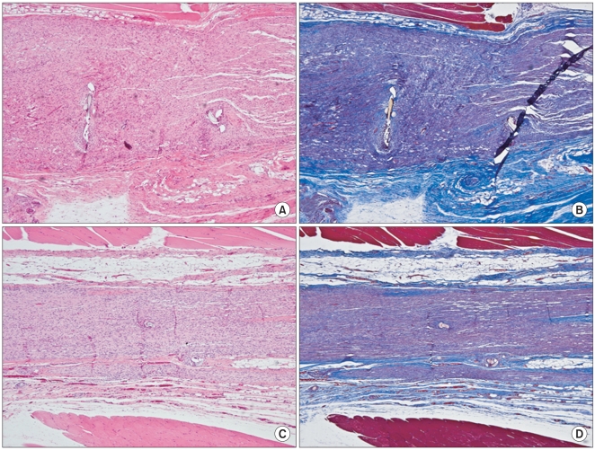
Fig. 9.
Photomicrographs showing the longitudinal sections of the sciatic nerves following cut and repair with saline (A, B) and with the hyaluronic acid-carboxymethylcellulose (HA-CMC) solution (C, D) at 12 weeks after surgery. The sections were stained with H&E (A, C) and with Masson's trichrome (B, D). Original magnification was × 40. In the nerves treated with saline, a mild inflammatory cell infiltration and a mostly parallel appearance of axons were observed at the repair site (A). However, in the nerves treated with the HA-CMC solution, a minimal inflammatory cell infiltration and a normal looking appearance of axons were demonstrated at the repair site (C). Compared to the HA-CMC solution group, the marked deposition of collagen surrounding the nerve was demonstrated in the saline group (B, D).

