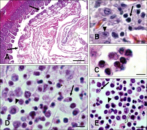
Fig. 1.
Histopathological microphotographs of apoptosis in new type gosling viral enteritis virus (NGVEV)-infected gosling. (A) Catarrhal hemorrhagic fibrinonecrotic enteritis with coagulative obstruction (arrows) was observed in the intestine tract of NGVEV-infected gosling that died 20 days post-infection (PI). (B) Apoptotic lymphocytes (arrow) and intestinal cells (arrowhead) were found in the small intestine. (C) Apoptotic cells and apoptotic bodies were identified in the intestinal. (D) Pyknosis (arrow) and necrosis (arrowhead) of the bursa of Fabricius (BF) lymphocytes. (E) Karyorrhexis (arrow), pyknosis (arrowshead), and necrosis (white arrow) of thymic lymphocytes were observed. H&E stain. Scale bars = 200 µm (A), 7 µm (B~D), 6 µm (E).

