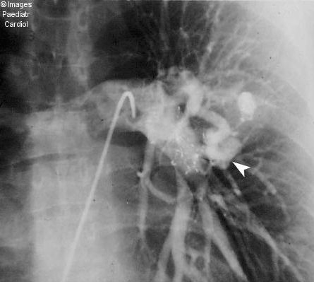
Figure 1(c).

Anteroposterior projection of a left pulmonary artery injection after placement of both devices demonstrates absence of flow within the large PAVM (compare with Fig 1a). The contrast enhanced smaller superimposed lesion (arrowhead) becomes apparent due to absence of contrast in the large PAVM
