
Figure 5(a).
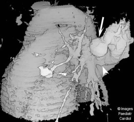
Surface shaded display reconstructed image from a set of contrast enhanced spiral CT slices demonstrates the huge PAVM (arrow) and its large feeding artery (arrowhead), as well as the two smaller lesions
Official websites use .gov
A
.gov website belongs to an official
government organization in the United States.
Secure .gov websites use HTTPS
A lock (
) or https:// means you've safely
connected to the .gov website. Share sensitive
information only on official, secure websites.
Surface shaded display reconstructed image from a set of contrast enhanced spiral CT slices demonstrates the huge PAVM (arrow) and its large feeding artery (arrowhead), as well as the two smaller lesions