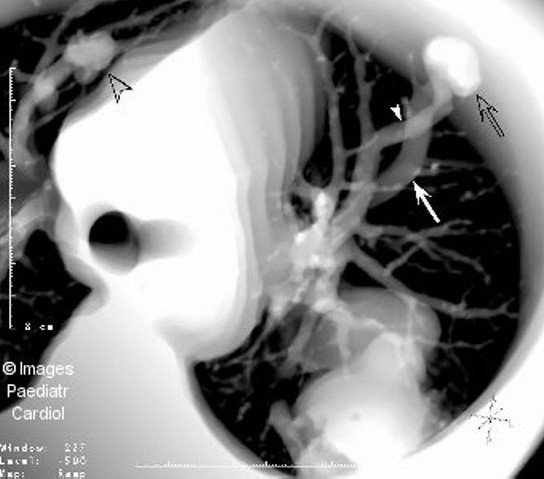
Figure 5(b).

Thick section multiplanar reformatted image from a set of contrast enhanced spiral CT slices shows part of the huge PAVM, as well as a small PAVM in the right upper lobe (open arrowhead). A smaller PAVM (open arrow) with its feeding artery (arrowhead) and dilated draining vein (arrow) is demonstrated in the left lung
