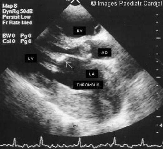
Figure 7.

Two-dimensional parasternal long-axis view of a patient with mitral stenosis, showing thickened valve cusps (arrow), with poor leaflet separation in diastole. Left atrium is enlarged, with a thrombus in the posterior aspect of it. Aortic valve is also stenotic
