Figure 14.

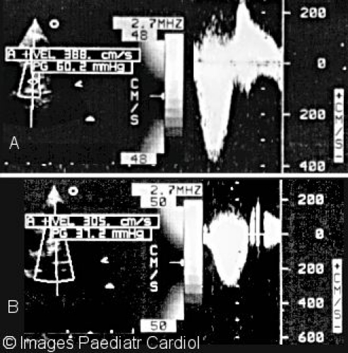
Continuous wave Doppler of right ventricular outflow tract and LPA showing A. Peak gradient before (60 mmHg) and B. Peak gradient after (37 mmHg) stenting

Continuous wave Doppler of right ventricular outflow tract and LPA showing A. Peak gradient before (60 mmHg) and B. Peak gradient after (37 mmHg) stenting