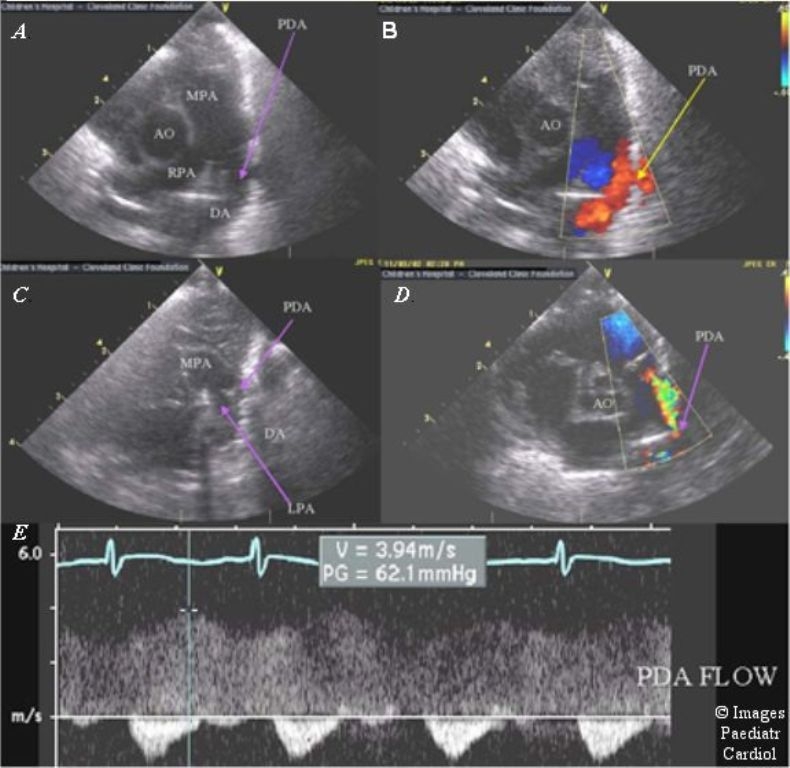
Figure 3.

Echocardiography of the PDA. The ductus can be well visualized from the left parasternal area (A) with low velocity flow back into the pulmonary artery from the aorta (B). After therapy with indomethacin the PDA significantly decreases in size (C) with aliasing color Doppler flow in a smaller jet (D), and a high velocity, restrictive spectral Doppler pattern (E). (MPA = main pulmonary artery, RPA = right pulmonary artery, Ao = aorta, PDA = patent ductus arteriosus, DA = descending aorta, LPA = left pulmonary artery)
