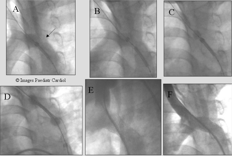
Figure 8.

Recurrent coarctation after subclavian flap aortoplasty (arrow). Note the proximity to the left carotid artery. Multiple LV angiograms obtained to accurately place the stent before and after uncovering the stent. The two ends of the balloon were allowed to expand slightly (C & D) with further imaging prior to fully deploying the stent (Max, IT Stent).
