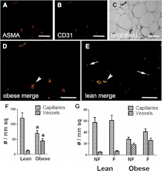
Fig. 1.
Identification of blood vessels in lean and obese adipose tissue. Obese (A–D) and lean (E) adipose tissue samples were immunoreacted with antibodies to CD31 (B, green) to identify endothelial cells and ASMA (A, red) to delineate the vessel wall, overlaid in D. C, The bright-field overlay of the same image. In the merged images from a representative obese (D) and lean (E) subject, larger blood vessels (arrowheads) are recognizable because they accumulate both ASMA and CD31. Capillaries are small and display only endothelial cell staining (arrows). Magnification, ×200; bar, 125 μm. F, Images from obese and lean subjects were scored for the number of capillaries and larger vessels, as described in Subjects and Methods, and normalized to cross-sectional area. Vessels are defined as structures that stained with CD31 and ASMA, and capillaries stained for only CD31. G, The areas containing the capillaries and vessels were categorized as fibrotic and nonfibrotic. *, P < 0.05 vs. lean.

