
Fig. 2.
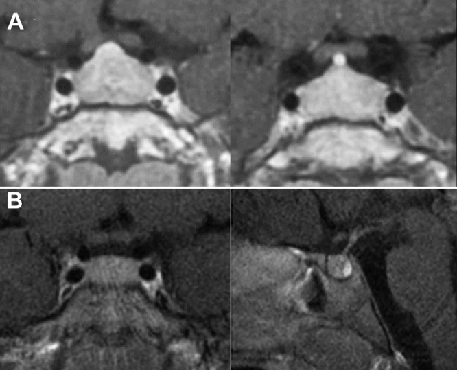
Preoperative contrast-enhanced MR imaging scans of the older brother (case 1) demonstrating symmetric enlargement of the pituitary (A) and the younger brother (case 3) demonstrating a slightly enlarged, symmetric gland (B). The posterior lobe is prominently seen. No focus suggestive of an adenoma is present in either scan.