Fig. 4.
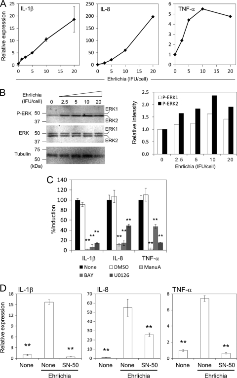
Induction of proinflammatory cytokines in THP-1 cells by E. chaffeensis is dependent on ERK and NF-κB pathways. (A) IL-1β, IL-8, and TNF-α induction in THP-1 cells by E. chaffeensis Wakulla was dose dependent as determined by real-time RT-PCR. Bacteria were added at different IFU/cell and incubated at 37°C for 2 h. (B) Phosphorylation of ERK in THP-1 cells treated with E. chaffeensis Wakulla was dose dependent. The titer of bacteria is the same as in panel A. Densities of phosphorylated ERK1 and ERK2 relative to those at 0 h as determined by densitometric analysis are shown on the right. (C) Effects of Ras, Raf, and MEK inhibitors on cytokine induction in THP-1 cells by E. chaffeensis Wakulla at 20 IFU/well. All inhibitors were added to the cell culture 45 min prior to infection and were present during infection. Final concentrations were 10 μM for manumycin A (ManuA, Ras inhibitor), 10 nM for U0126 (MEK inhibitor), 250 nM for BAY43-9006 (BAY, Raf-1 inhibitor). DMSO (1%) was added as a negative control. Data shown are means and standard deviations from triplicate assays. **, significantly different from either the nontreated or DMSO-treated control (ANOVA, P < 0.01). (D) THP-1 cells were incubated with or without E. chaffeensis Wakulla at 20 IFU/cell for 2 h in the presence or absence of 100 μg/ml SN-50, a cell-permeable NF-κB inhibitory peptide. Expression of cytokine genes was determined by quantitative RT-PCR. Data shown are means and standard deviations from triplicate assays. **, significantly different from Ehrlichia, nontreated control (ANOVA, P < 0.01). Data are representative of results of at least three independent experiments.

