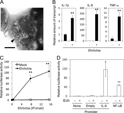
Fig. 5.
E. chaffeensis induces cytokines and activates IL-8 and NF-κB promoters in HEK293 cells. (A) HEK293 cells infected with E. chaffeensis. Host-free E. chaffeensis was inoculated and incubated with HEK293 cells for 6 days. The cells were stained with Diff-Quik. An arrowhead indicates multiple morulae of E. chaffeensis in the cytoplasm of a HEK293 cell. Bar = 5 μm. (B) IL-1β, IL-8, and TNF-α induction in HEK293 cells 2 h after exposure to E. chaffeensis Wakulla. Bacteria were added at 40 IFU/cell and incubated at 37°C for 2 h. Quantitative RT-PCR. (C) Dose-dependent activation of the IL-8 promoter. Luciferase reporter activity of pK666-transfected HEK293 cells was determined after incubation with bacteria at 0 to 15 IFU/cell or corresponding uninfected DH82 cell extract control (Mock) at 37°C for 2 h. **, significantly different from control (Student's t test, P < 0.01). (D) Luciferase activity of HEK293 cells transfected with pGL4.17 (Empty, vector harboring promoterless luciferase), pK666 (IL-8, IL-8-luciferase), pGL4.32 (NF-κB, NF-κB responsive element-luciferase) or none 2 h after incubation with E. chaffeensis Wakulla (Ech +) at 15 IFU/cell or corresponding uninfected DH82 cells (Ech −) at 37°C. Data shown are means and standard deviations from triplicate assays. * and **, significantly different from no or empty plasmid control (Student's t test; P < 0.05 and P < 0.01), respectively. Data are representative of results of at least three independent experiments.

