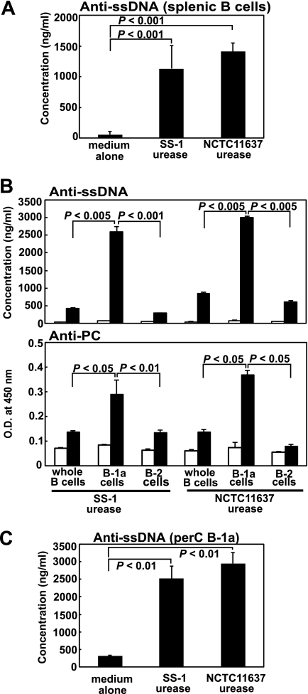
Fig. 1.
Autoantibody secretion from purified murine splenic B cells as well as their sorted B-1 or B-2 cells stimulated with purified H. pylori urease. (A) Purified B cells (1 × 10⁶) were cultured with 10 μg/ml purified H. pylori urease from either the SS-1 or NCTC 11637 isolate for 7 days, and the supernatants were harvested to measure their anti-ssDNA autoantibody production by ELISA. Data are means ± SDs (n = 6). (B) Either 2 × 10⁵ purified B cells or the same number of their sorted B-1a or B-2 cells were cultured with 10 μg/ml purified H. pylori urease (filled columns) or PBS (open columns) for 7 days in CM, and the supernatants were harvested to measure either their anti-ssDNA (top) or anti-PC (bottom) autoantibody production. Data are means ± SDs (n = 6). O.D., optical density. (C) Purified peritoneal cavity-derived B-1a (perC B-1a) cells (1 × 10⁵) were cultured with 10 μg/ml purified H. pylori urease for 7 days, and the supernatants were harvested to measure their anti-ssDNA autoantibody production. Statistical significance was determined by the Student t test, and the values are given in each graph.

