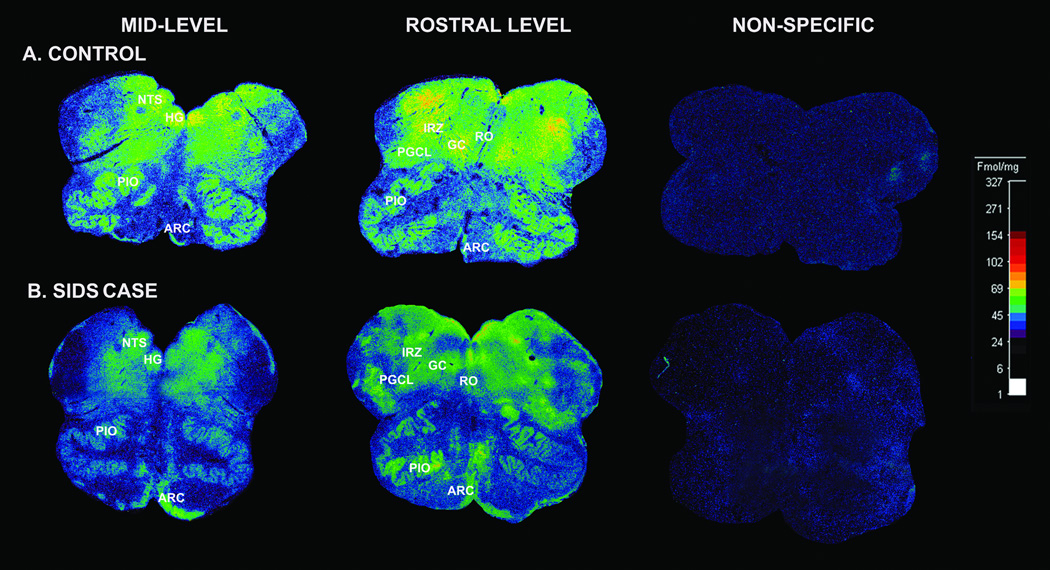
Figure 2.
γ-Aminobutyric acidA (GABAA) receptor binding in the medulla and cerebellum. (A, B) Binding in the medullary regions is lower in the 44 postconceptional weeks SIDS case (B) vs. the 44 postconceptional weeks control (A), except in the arcuate nucleus in which the binding levels are similar. Fragments of cerebellar tissue (dotted lines) serve as an internal control of receptor binding levels. There is no difference between the cases in binding levels in the cerebellar tissue. ARC, arcuate nucleus; HG, hypoglossal nucleus; NTS, nucleus of the solitary tract; PIO, principal inferior olive; RO, raphé obscurus.

