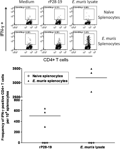
Fig. 7.
Outer membrane protein P28-19-specific CD4+ T cells develop during E. muris infection. We determined the frequencies of P28-19-specific IFN-γ-producing CD4+ T cells in the spleens of mice infected with E. muris by flow cytometry. The spleens of mice infected with E. muris had higher frequencies of P28-19-specific IFN-γ-producing CD4+ T cells on day 45 after infection than the spleens of naïve uninfected mice. The frequencies of E. muris-specific IFN-γ-producing CD4+ T cells in the spleens of the same mice detected following in vitro stimulation with the E. muris whole-cell lysate were shown for comparison. In the graph at the bottom of the figure, each symbol represents the value for an individual mouse, and the short horizontal lines represent the mean for the group.

