Fig. 4.
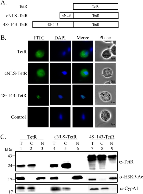
Region in Myb2 sufficient for nuclear import of a TetR fusion protein. (A) The region spanning aa 48 to 143 of Myb2 or the cNLS from the SV40-T antigen was fused with a C-terminal bacterial tetracycline repressor, TetR. (B) Subcellular localization of TetR, cNLS-TetR, and 48∼143-TeR was examined by IFA with the anti-TetR antibody. The signal was shown as green fluorescence (FITC). The nucleus was stained with DAPI, and cell morphology was recorded by phase-contrast microscopy. Bar, 5 μm. (C) Total lysates (T) from cells overexpressing TetR, cNLS-TetR, and 48∼143-TeR were fractionated into the nuclear (N) and cytosolic (C) fractions. Protein samples were separated in 12% gel for Western blotting using the anti-TetR antibody. Duplicate blots were examined by the anti-acetyl-histone H3(Lys9) (α-H3K9-Ac) and anti-CypA1 (α-CypA1) antibodies.

