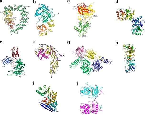
Fig. 3.
Three-dimensional structures of 10 E. coli PG hydrolases. (a) Slt70. (Reprinted from reference 276 with permission of Elsevier.) (b) MltA. (Reprinted from reference 289 with permission of Elsevier.) (c) MltB. (Reprinted from reference 277 with permission of Elsevier.) (d) MltE. (Reprinted from reference 3 with permission from the publisher. Copyright 2011 American Chemical Society.) (e) PBP4. (Reprinted from reference 125 with permission from the publisher. Copyright 2006 American Chemical Society.) (f) PBP5. (Reprinted from reference 46 with permission of the publisher.) (g) PBP6. (Reprinted from reference 36 with permission from the publisher. Copyright 2009 American Chemical Society.) (h) MepA. (Reprinted from reference 161 with permission of the publisher.) (i) YcjG epimerase. (Reprinted from reference 84 with permission of the publisher. Copyright 2001 American Chemical Society.) (j) AmiD dimer. (Reprinted from reference 124 with permission of Elsevier.)

