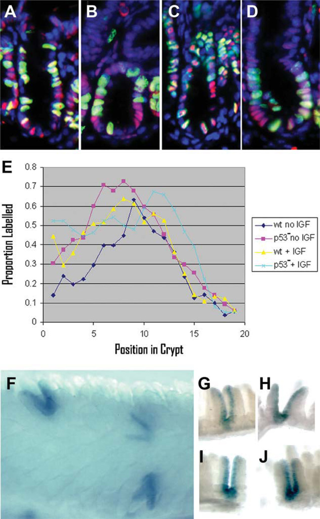
Figure 4.
Pulse labeling and Mcm2-CreERT2-mediated marking studies confirm an increased rate of cycling in the stem cells of p53 null mice relative to wild type. In panels (A–E) wt or p53 null mice were treated with daily IGF1 injections for 3 days and compared to mice of the same genotypes that had not been treated with IGF1. A pulse of CldU was given 2 hours prior to harvesting the small intestine. Panels (A–D) show 7-µm-thick longitudinal paraffin sections stained for Mcm2 (red), CldU (green), and DAPI (blue) where panel (A) is wt, no IGF1; panel (B) is p53 null, no IGF1; panel (C) is wt, IGF1 treated; and panel (D) is p53 null, IGF1 treated. Panel (E) shows of the proportion of Mcm2 stained nuclei that are also stained for CldU (y-axis) plotted as a function of position of the nuclei from the base of the crypt (x-axis) from 50 or more crypts for each of the different experimental conditions as indicated in the box. Panel (F) is a bright-field image of a whole mount preparation of small intestine stained for β-galactosidase activity from a p53 null, Mcm2-CreERT2 mouse carrying a Cre-dependent lacZ reporter at approximately 1 month following tamoxifen treatment. Panels (G–J) are individual β-galactosidase expressing crypts prepared by microdissection. Abbreviation: IGF, insulin-like growth factor 1.

