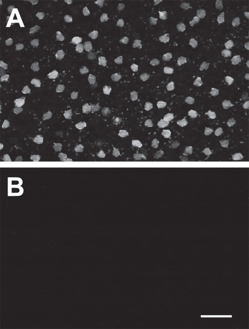
Figure 1.
Cross-signal contamination was very low. Flat-mounted retina labeled with anti-parvalbumin antibody and visualized with Alexa Fluor 488-conjugated secondary antibody, as described for Figures 4, 5, and 6. (A) Normal image optimized to capture the signal generated by the 488 dye. (B) The exact same field obtained with 488 illumination but using the settings and filter sets normally used to acquire the image generated by secondary antibodies conjugated to cy3, Alexa Fluor 555 or Alexa Fluor 594. Scale bar is 25 µm. No cross-signal contamination is visible at these settings. If brightness and contrast are greatly increased, a signal on the order of 1% of panel A can be seen (not shown). Images of all double-labeled preparations were used for data analysis only if cross-signal contamination was as low as shown here.

