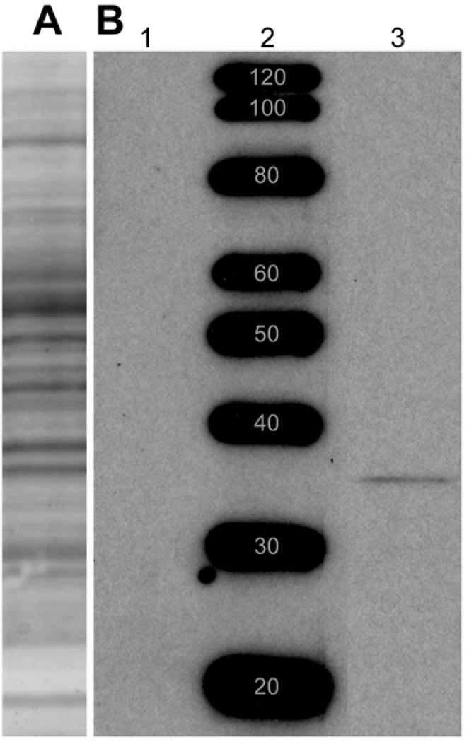
Figure 2.
Western Blot: 100-µg samples of rat retina lysate and protein standards were resolved by electrophoresis and transferred onto nitrocellulose. (A) Ponceau S probe of a single lane of retina lysate shows that multiple protein bands transferred from the gel to the nitrocellulose. The positions of molecular weight standard proteins run adjacent to this lane are identical to those in B and, therefore, not shown. (B) Specific DARPP-32 staining: Retina lysate samples were run in lanes 1 and 3, on either side of a lane of molecular weight standard proteins. Lane 3 was probed with anti-DARPP-32 antibody, while lane 1 was probed with anti-DARPP-32 antibody that had been pre-incubated overnight with blocking peptide. Anti-DARPP-32 antibody stained a well-focused protein band in lane 3, and this staining was blocked completely by the antibody blocking peptide (lane 1). The molecular weight of this band was estimated to be 32 kD, by comparison with a standard curve constructed from the distances migrated by the protein standards (lane 2). Note that the blot in B was prepared from a different gel than the blot in A. Blot in A included to show typical range of proteins obtained from retinal homogenates. Bands in B, lane 2, are prominent because Magic Mark standards have been optimized for visualization with HRP.

