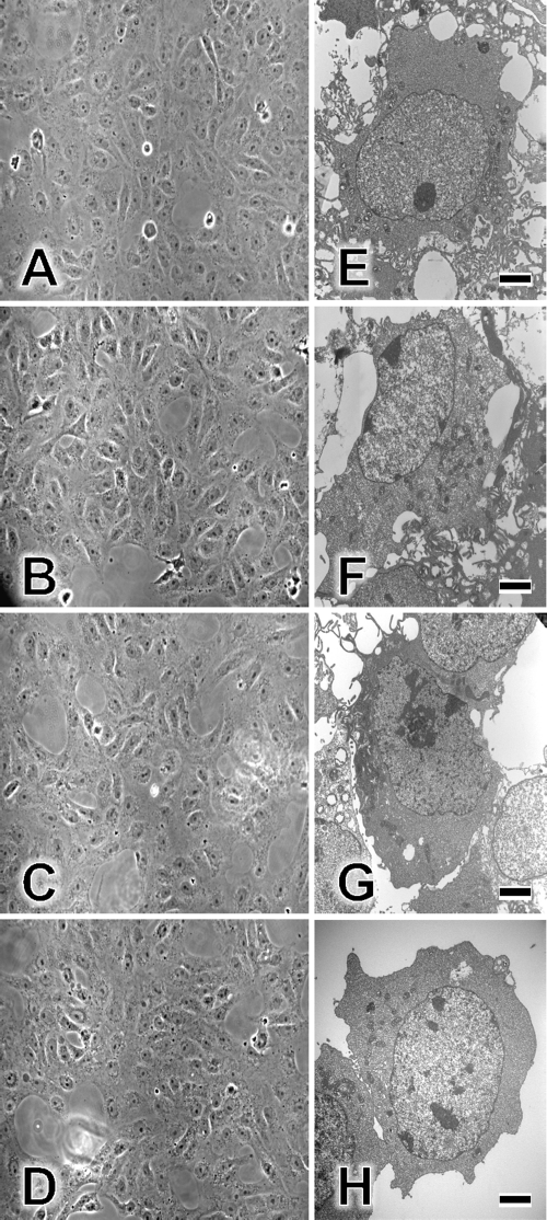
Fig. 5.
Effects of TF-3, theasinensin A, and EGCG on Vero cells. Vero cell monolayers were incubated for 1 h at 37°C in tissue culture medium with 1% FCS (A), medium with 100 μM EGCG (B), medium with 100 μM theasinensin A (C), or medium with 100 μM TF-3 (D). Monolayers were then examined by phase-contrast microscopy and photographed at ×20 magnification. Individual Vero cells were examined by EM as described in Materials and Methods. EGCG and dimers were utilized at 100 μM. (E) Control cells; (F) EGCG-treated cells; (G) theasinensin A-treated cells; (H) TF-3-treated cells. Scale bars are 2 μm (panels E to H).

