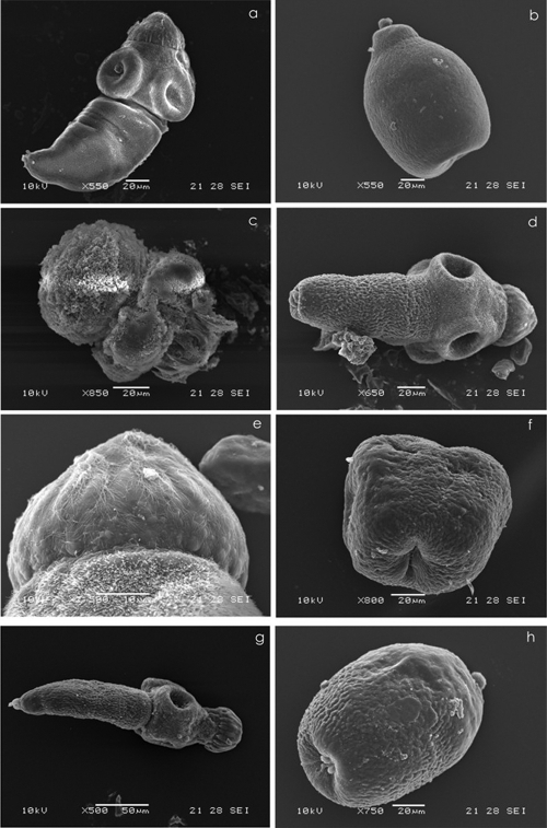
Fig. 2.
Representative images of the scanning electron microscopy (SEM) of protoscoleces cultured in vitro for 6 days in the presence of medium containing methanol (10 μl) (control group) or FLBZ, R-FLBZ, ABZ, or ABZSO. (a) Evaginated control protoscolex. (b) Invaginated control protoscolex. (c) Evaginated protoscolex cultured with FLBZ. Note the extensive drug-induced damage with contraction in the soma region, the tegument being markedly altered and also shedding microtriches, and disorder on the rostellum. (d, e, and f) Evaginated protoscolex cultured with R-FLBZ. The altered tegument of the soma region and loss of microtriches on the rostellum can be observed. Details of the rostellum from protoscolex are shown in panel d. and invaginated protoscolex with an extensive damage affecting the tegument. (g) Evaginated altered protoscolex in the presence of ABZ. The shedding of microtriches can be observed in the scolex region either on suckers or on the rostellum. (h) Invaginated protoscolex, clearly altered after culture in the presence of ABZSO.

