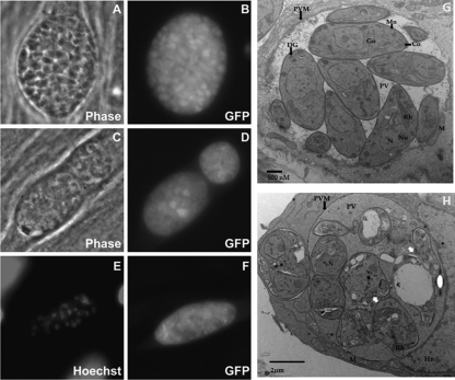
Fig. 7.
Phenotypic effects of F1792-0016 on parasites in vitro. (A to F) Parasites were treated with vehicle (A and B) or 2 μM F1792-0016 (C to F), fixed, and observed using fluorescence microscopy and electron microscopy. (A and B) Parasites treated with vehicle for 48 h. (C and D) Parasites treated for 72 h with F1792-0016 exhibit significant vacuole disruption that is evident both in phase-contrast and in the GFP channel. (E and F) Nuclear staining and GFP fluorescence of a disrupted vacuole from parasites treated for 48 h with F1792-0016. (G and H) Parasite ultrastructure observed using transmission electron microscopy. (G) Vehicle-treated cells. (H) F1796-0016 causes disruption of the parasite plasma membrane (white arrows), and the PV is more electron dense than in the controls, possibly containing the contents of lysed parasites. Co, conoid; DG, dense granules; Go, Golgi complex; Hn, host nucleus; M, host mitochondria; Mn, micronemes; N, nucleus, Nu, nucleolus; PVM, parasitophorous vacuolar membrane; PV, parasitophorous vacuole; Rh, rhoptries. Magnifications, ×15,000 (G) and ×12,000 (H).

