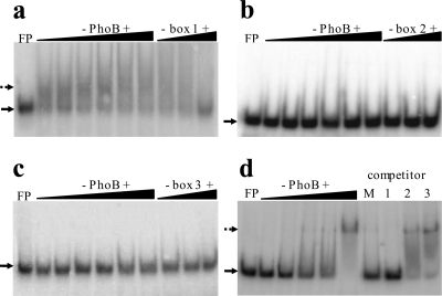
Fig. 3.
Binding of PhoB to individual Pho boxes in the phoBR regulatory region. Fifty fmol of radiolabeled double-stranded oligonucleotides corresponding to each one of the three Pho boxes was used in EMSAs with increasing PhoB concentrations. The binding patterns (dotted arrows) of Pho box 1 (a), Pho box 2 (b), and Pho box 3 (c) to PhoB at 2, 5, 10, 20, 30, and 37 μM are presented. Competition assays were carried out with 37 μM PhoB plus 50, 250, and 500 fmol of competing nonlabeled oligonucleotides. (d) The labeled oligonucleotide corresponding to Pho box 1 was incubated with increasing PhoB concentrations (0.5, 2, 5, 10, and 30 μM). Competition assay was performed with PhoB at 30 μM against 500 fmol of competing nonlabeled Pho boxes containing fragments. Lane M, equimolar mixture of Pho boxes 1, 2, and 3 (a total of 500 fmol); lane 1, 500 fmol of Pho box 1; lane 2, 500 fmol of Pho box 2; lane 3, 500 fmol of Pho box 3. FP, free probe (solid arrows).

