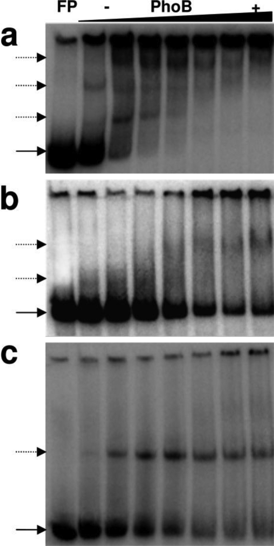
Fig. 4.
Binding patterns of PhoB to three distinct phoBR promoter fragments. Fifty fmol of labeled double-stranded probes were used with increasing PhoB concentrations (0, 0.5, 2.5, 5, 10, 20, 30, and 37 μM). Binding patterns of probes containing boxes 1, 2, and 3 (a), boxes 2 and 3 (b), or box 1 alone (c) are shown. In the first lane, the free probe (FP) position is indicated by a solid arrow; dotted arrows point to shifted bands.

