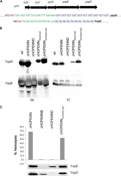
Fig. 2.
(A) Location of yopD on the translocator operon on the pYV plasmid. DNA and amino acid sequence of the Strep tag and the 10-amino-acid linker. The +2 codon corresponding to an asparagine is depicted in red, the Strep tag is in green, and the 10-amino-acid linker is in blue. (B) Immunoblots of anti-YopB and anti-YopD from total cell (TC) and supernatant (SN) of bacterial cultures shifted for 4 h at 37°C to induce secretion. (C) Lytic activity on red blood cells after four periods of 10 min of contact with Y. enterocolitica ΔHOPEMN, ΔHOPEMNB, ΔHOPEMND, and ΔHOPEMN-AsnStrep-YopD bacteria. One hundred percent hemolysis corresponds to RBCs lysed with 0.1% Triton X-100. The immunoblotting of anti-YopB and anti-YopD was performed on the floated infected RBC membranes.

