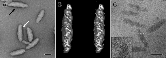
Fig. 2.
High-resolution electron micrograph, tomographic reconstruction of a negatively stained chlorosome, and cryo-electron microscopic images of chlorosomes of “Ca. Chloracidobacterium thermophilum.” (A) Negatively stained chlorosomes. The black arrow points to the ridges and grooves visible on one of the surfaces of the chlorosomes. The white arrow points to the flattened surface area that presumably includes the CsmA-containing baseplate structure. (B) Stereo image of a negatively stained chlorosome as an iso-surface representation from a tomographic reconstruction. The internal domains described in Results are indicated by the dashed ellipses. (C) Cryo-electron microscopic image of isolated chlorosomes embedded in vitreous ice. Note the twisted or folded lamellae. (Insert) Enlarged view of the boxed area, showing the curved lamellae with 2.3-nm spacing. The lamellar spacing was determined from a Fourier transformation of the data (see references 20 and 45 for further details). Bars, 50 nm.

