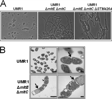
Fig. 6.
Morphological characterization of mltE and mltC mutants. (A) Light microscopy of the mltE mltC and mltE mltC STM4264 mutants. The mltE mltC double mutant and the mltE mltC STM4264 triple mutant display the formation of long chains when grown at 28°C in LB without salt medium. The wild-type UMR1 shows rod-shaped cells of standard length. Magnification, ×630. (B) Transmission electron microscopy (TEM) of the ΔmltE ΔmltC double mutant. Long chains of cells are observed in the ΔmltE ΔmltC double mutant due to an impairment of the cleavage of the PG septum (arrow). The wild-type UMR1 shows rod-shaped cells of 0.8 to 1 μm standard length without impairment in the cleavage of the PG septum. Bars indicate 1 (left panel) or 0.2 μm (right panel).

