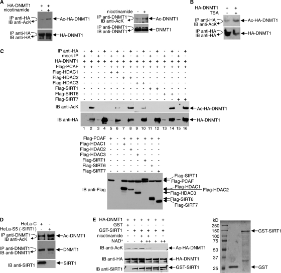
Fig. 2.
Deacetylation of DNMT1 by SIRT1 in vivo and in vitro. (A) Anti-HA (left) or anti-DNMT1 (right) immunoprecipitates from 293T cells, which were transfected with HA-DNMT1 expression plasmid (left panel) or untransfected (right panel) and treated with 15 mM nicotinamide for 12 h, were immunoblotted with antibody to AcK. The membranes were stripped and reprobed with anti-HA or anti-DNMT1. (B) 293T cells expressing HA-DNMT1 received 400 ng/ml of TSA for 2 h. Anti-HA immunoprecipitates were immunoblotted with antibody to acetyllysine or HA. (C) 293T cells were transfected with HA-DNMT1, Flag-PCAF, and Flag-HDAC1, -2, or -3 or Flag-SIRT1, -6, or -7 as indicated. Cell lysates were immunoprecipitated with antibody to HA or were mock precipitated (IgG), and immunoprecipitates were immunoblotted (IB) with antibody to acetyllysine. The membrane was stripped and reprobed with anti-HA. A separate Western blot assay was performed with anti-Flag antibody to assess Flag-HDAC and Flag-SIRT expressions (bottom). (D) HeLa cells were transfected with either shRNA pSuper-SIRT1 (HeLa-S) or scrambled control shRNA (HeLa-C) and grown in 1 μg/ml puromycin for 2 weeks. SIRT1 depletion in HeLa-S cells was assessed by Western blot assays, and one colony (HeLa-S5) was selected for further analysis. Anti-DNMT1 immunoprecipitates were immunoblotted with antibodies to acetyllysine or DNMT1. Immunoblot assays were also performed to assess SIRT1 expression. (E) (Left) HA-DNMT1 was first hyperacetylated in vivo by coexpression with PCAF in 293T cells, and then cell lysates were prepared and immunoprecipitated with antibody to HA. In vitro deacetylation reactions were performed by incubating anti-HA immunoprecipitates (Ac-HA-DNMT1), 1 μg GST or GST-SIRT1, NAD+, and 10 mM nicotinamide as indicated. Reaction products were immunoblotted with antibody to acetyllysine. The membrane was stripped and reprobed with anti-HA or anti-SIRT1. (Right) The quality of bacterially expressed, purified GST and GST-SIRT1 proteins was assessed by SDS-PAGE and Coomassie blue staining.

