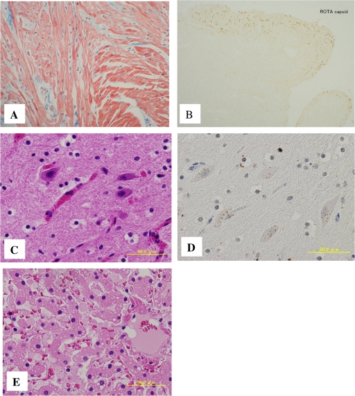
Fig. 1.
Pathological findings in heart, brain, and liver tissue. (A) Hypertrophic cardiomyopathy with focal disorganized myocardial architecture. (B) Endocardium showing immunohistochemical detection of rotavirus capsid protein with a mouse monoclonal antibody. (C) Brain tissue with hematoxylin-and-eosin staining. (D) Brain tissue stained with anti-VP6 antibody. The brown color indicates a positive reaction. (E) Liver section with hematoxylin-and-eosin staining showing a multitude of small lipid droplets.

