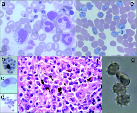
Fig. 1.
Malaria diagnosis and characterization of the clinical isolate. (a to d) Giemsa-stained blood smears from the patient showing mostly ring and trophozoite forms of Plasmodium falciparum (a), but schizonts (b), gametocytes (c), and merozoites (d) were also observed. (e) Smear of the culture from the patient isolate showed optimal growth at high parasitemias. (f) Spleen slides (hematoxylin and eosin) showing local deposition of the malaria pigment hemozoin in macrophages. (g) Rosette formation by the P. falciparum isolate.

