Fig. 8.
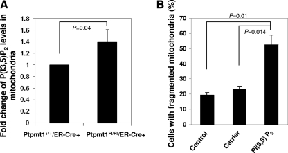
PI(3,5)P2 is accumulated in the mitochondria in Ptpmt1-depleted cells, and overloading of PI(3,5)P2 results in defective mitochondrial dynamics. (A) 4-OHT-treated Ptpmt1+/+/ER-Cre+ and Ptpmt1flox/flox/ER-Cre+ MEFs were transfected with Mito-DsRed2 and then immunostained with anti-PI(3,5)P2 antibody. PI(3,5)P2 was visualized using Alexa Fluor 488-conjugated anti-mouse secondary antibody. Green fluorescence intensity in Mito-DsRed2-positive areas in Ptpmt1flox/flox/ER-Cre+ cells (n = 137) was quantified and normalized against that in the Ptpmt1+/+/ER-Cre+ control (n = 131) using laser scanning cytometric analyses. Results shown are means ± standard deviations of three independent experiments. (B) PI(3,5)P2 (di-C16) (1 μM) was delivered (shuttled) into Ptpmt1+/+/ER-Cre+ MEFs using Shuttle PIP kits following the protocol provided by the manufacturer (Echelon Biosciences, Inc.). Carrier 2 was used to deliver PI(3,5)P2. Cells were stained with Mitotracker Red 4 h later. The percentage of the cells with fragmented mitochondria was determined. Representative results of one experiment (average of three different fields, and 200 cells were counted for each field) are shown. Experiments were repeated using two different cell clones, and similar results were obtained.

