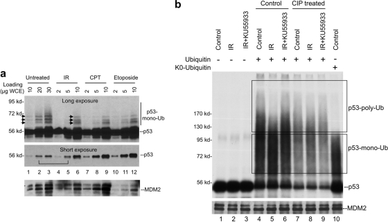
Fig. 1.
DNA damage inhibits p53 ubiquitination. (a) U2OS cells were treated with 10 Gy of IR for 4 h, 0.5 μM CPT for 8 h, or 50 μM etoposide for 8 h. Cells were suspended in Laemmli SDS-PAGE sample buffer with 10 mM iodoacetamide and immediately boiled. Whole-cell lysate was loaded at the indicated amounts and analyzed by p53 Western blotting. Long and short exposures were obtained to determine the levels of p53-ubiquitin conjugate and unmodified p53. (b) SJSA cells were treated with 10 Gy of IR for 2 h in the presence or absence of ATM inhibitor KU55933. MDM2 was immunoprecipitated by using 2A9 antibody. The MDM2-loaded beads were then used to capture in vitro-translated p53, followed by incubation with E1, E2, and ubiquitin. p53 ubiquitination was detected by autofluorography. Dephosphorylation treatment using CIP was performed on MDM2 beads prior to the capture of p53. p53 monoubiquitination products were identified by the use of K0-ubiquitin in the ubiquitination reaction.

