Fig. 5.
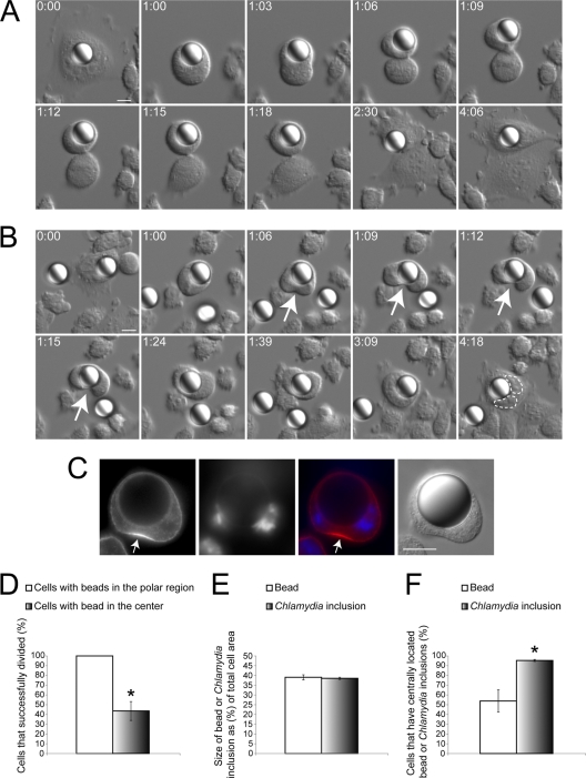
Centrally located large polystyrene beads can recapitulate the unilateral cleavage furrow defect. (A and B) CHO-IIA cells that internalized a bead were subject to DIC imaging. Numbers indicate hours:minutes. Scale bars, 10 μm. (A) Cells with beads in the polar region were able to form bilateral cleavage furrows and successfully divide into two discrete daughter cells (n = 16 out of 16). (B) When the bead was in the cell center, a unilateral cleavage furrow (arrows) could be induced. Cytoplasmic division frequently failed in cells with centrally positioned beads (n = 18 out of 27). Two nuclei were outlined with dashed lines. (C) RhoA (red) only accumulated at the equatorial cell cortex away from the centrally located latex bead in CHO IIA cells. DNA was stained with DAPI (blue) (n = 10 out of 10). (D) Cells containing centrally located beads had a significantly lower success rate of division than cells containing beads in the polar region (P < 0.005). (E) The relative size of 15-μm beads compared to host cells was indistinguishable from that of the Chlamydia inclusions at 36 hpi (P > 0.74). (F) Chlamydia inclusions localized to the cell equator significantly more often than similarly sized beads during telophase (P < 0.005). (D to F) Error bars represent the SEM from at least three independent experiments.

