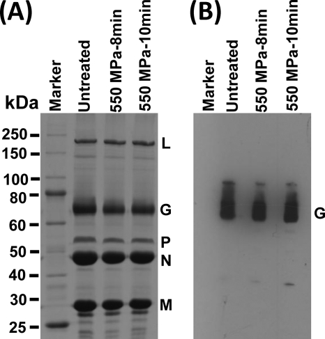
Fig. 6.
The effect of HPP on VSV proteins. (A) Visualization of VSV structural proteins by 12% SDS-PAGE. Purified VSV was treated at 550 MPa and 20°C for 8 min and 10 min, respectively. Ten micrograms of total viral proteins was separated by SDS-PAGE followed by Coomassie blue staining. (B) Western blot analysis of VSV G protein. The viral proteins of untreated and treated VSV samples were separated by SDS-PAGE and subjected to Western blotting using a monoclonal anti-VSV G protein.

