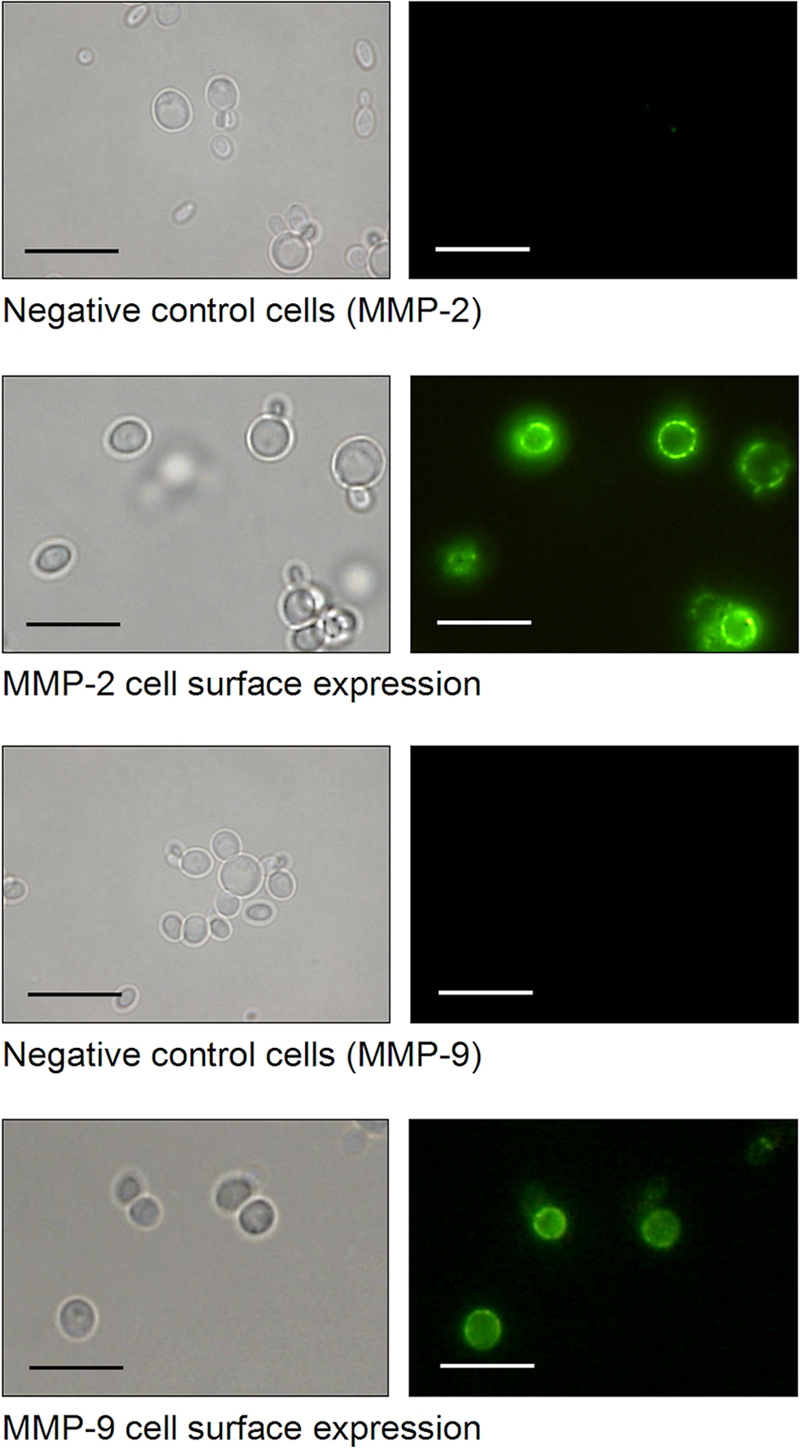
Fig. 3.

P. pastoris cell surface display of human MMP-2 and MMP-9 verified by indirect immunofluorescence microscopy. Cells of P. pastoris KM71 expressing the indicated MMP were cultivated for 72 h under inducing conditions in the presence of methanol. The MMPs were labeled with anti-MMP antibodies and fluorescein isothiocyanate (FITC)-conjugated anti-rabbit IgG and analyzed by fluorescence microscopy (Keyence BZ-8000; λA = 480 nm; λE > 510 nm). Scale bars = 10 μm.
