Fig. 4.
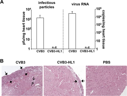
Cardiotropism and cardiopathogenicity of CVB3-HL1 in vivo. (A) CVB3-HL1 lacks cardiotropism in vivo. Nine BALB/c mice were infected with 5 × 10⁵ PFU of CVB3 or CVB-HL1. As a control, 6 mice were treated with PBS. Seven days after CVB3 infection, organs were harvested for virus detection and histopathological analysis. CVB3 positive-strand RNA and infectious virus numbers in the heart tissue were quantified by real-time RT-PCR and a standard plaque assay, respectively. Quantitative analysis showed a complete absence of CVB3-HL1 in the heart, whereas CVB3 RNA (right) and infectious CVB3 particles (left) were abundantly detected. n.d., not detectable. (B) Heart sections of CVB3- and CVB3-HL1-infected mice were stained with hematoxylin and eosin (magnification, 100-fold). CVB3-infected animals showed an expanded inflammation area at the pericardium (thick arrows) and some inflammatory spots with mononuclear cells in the myocardium (thin arrows). Heart tissues of CVB3-HL1-infected animals showed faint pericardial infiltrations with mononuclear cells.

