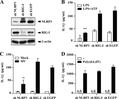
Fig. 2.
NLRP3 inflammasome activation by MV. (A) Expression levels of NLRP3 and RIG-I in PMA-stimulated THP-1 cells expressing shRNA targeting NLRP3, RIG-I, or EGFP mRNAs were examined by Western blot analysis at 24 h after LPS (1 μg/ml) stimulation. β-Actin was used as an internal control. (B to D) PMA-stimulated THP-1 cells expressing shRNA targeting mRNAs of NLRP3, RIG-I, or EGFP were treated with LPS (5 ng/ml) or LPS (5 ng/ml) plus ATP (5 mM) (B), MV (C), or poly(dA·dT) (400 ng/ml) (D). Cell-free supernatants were collected 24 h after treatment and analyzed for IL-1β by ELISA. n.d., not detected. Values are means and standard deviations from triplicate samples. The data shown are representative of at least three experiments. **, P < 0.01; ***, P < 0.001.

