
Fig. 3.
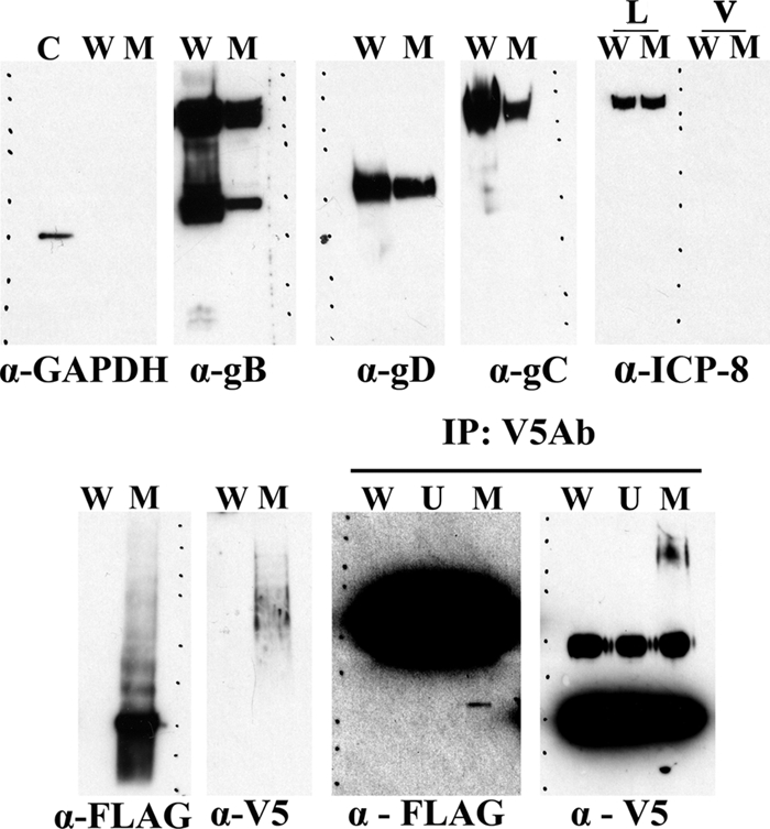
Detection of gK and UL20 on gradient-purified virions. Western immunoblot analysis of double-gradient-purified virions was performed using anti-V5 (gK) and anti-FLAG (UL20) antibodies. Individual lanes are labeled as follows: C, positive control for GAPDH; W, lysates from purified wild-type virus [HSV-1(F)-YE102]; M, lysates from purified mutant virus YE102-VC1; L, lysates from infected Vero cells; V, samples derived from purified virions. Individual blots were probed with anti-GAPDH, anti-ICP8, anti-gB, anti-gD, anti-gC, anti-FLAG (UL20), or anti-V5 (gK) antibody as indicated. The panel labeled at the top as IP:V5Ab shows immunoblots of immunoprecipitations with anti-V5 (gK) antibody probed with either anti-FLAG (UL20) or anti-V5 (gK) antibodies. In this panel, lanes are immunoprecipitates from mock-infected Vero cells (U), HSV-1(F)-YE102 purified virions (W), or YE102-VC1 purified virions (M). Molecular mass standards are shown with dots on each panel (250, 150, 100, 75, 50, 37, 25, 20, 15, and 10 kDa; Precision Plus protein standards; Bio-Rad). HRP-conjugated goat anti-mouse (HRP-GAb; IgG) was used for all data, except data shown on the panel labeled IP:V5Ab, wherein the F(ab)2- and Fc-purified portions of the HRP-GAb IgG were used for the α-V5 and α-FLAG panels, respectively.