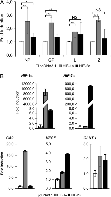
Fig. 4.
Effect of HIF-α subunit transfection on LCMV MX gene expression under normoxia. (A) HeLa-MX cells were transfected transiently with HIF-1α or HIF-2α expression vector and maintained under normoxia. Empty pcDNA3.1 was used to adjust the total DNA content in the control sample. Expression of viral genes was assessed 72 h after transfection using quantitative RT-PCR. Fold induction was determined in comparison with the values from pcDNA3.1-transfected cells. Values represent means from three separate experiments, each done in triplicates. Error bars denote the standard deviations. *, P < 0.05; **, P < 0.01; ***, P < 0.001; NS (not significant), P > 0.05. (B) Transfected HeLa-MX cells were analyzed by quantitative RT-PCR to determine expression levels of HIF-1α and HIF-2α subunits and their transcriptional targets. Data were analyzed and illustrated as described above.

